| 反应性 | H M R Mk |
| 灵敏度 | 内源性 |
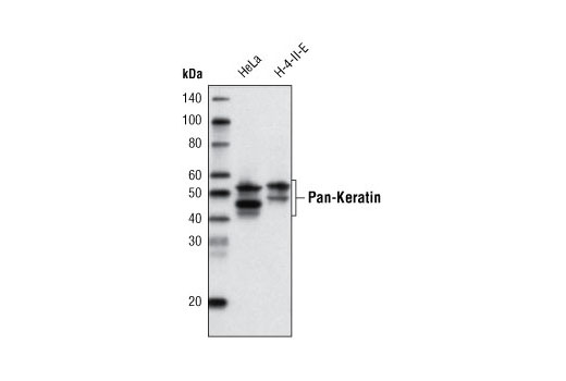
| MW (kDa) | 46-58 |
| 来源/同种型 | 小鼠 IgG1 |
产品信息
| MW (kDa) | 46-58 |
| 应用 | 稀释度 |
|---|---|
| 蛋白质印迹法 | 1:1000 |
蛋白质印迹法就是将膜与 5% w/v 脱脂奶粉、1X TBS 以及 0.1% Tween® 20 中的稀释的一抗在 4°C 下进行夜间孵育,同时轻轻晃动。
注意:请参阅一抗产品网页了解推荐的抗体稀释度。
注:使用反渗透去离子水 (RODI) 或同等级别的水配制溶液。
上样 20 µl 到 SDS-PAGE 凝胶 (10 cm x 10 cm) 上。
注意:建议将预染蛋白分子量标准品(#59329,10 µl/泳道)上样,以验证转膜的效率,生物素化蛋白质标准品(#7727,10 µl/泳道)可以直接在膜上显出条带以确定分子量。
注意:体积适用于 10 cm x 10 cm (100 cm2) 的膜;对于不同尺寸的膜,可相应调整体积。
请勿加入用于检测生物素化蛋白标准品的 Anti-biotin, HRP-linked Antibody。没有必要。Streptavidin-HRP 也将会使生物素化标准品可视化。
* 避免反复接触皮肤。
发布时间 2005 年 6 月
修订时间 2020 年 6 月
实验步骤编号:265
人, 小鼠, 大鼠, 猴
使用 A-431 细胞的细胞骨架制备物对动物进行免疫接种来产生单克隆抗体。
角蛋白(细胞角蛋白)是主要在上皮细胞中表达的中间丝蛋白。Keratin heterodimers composed of an acidic keratin (or type I keratin, keratins K9-K28) and a basic keratin (or type II keratin, keratins K1-K8 and K71-K80) assemble to form filaments. Keratin isoforms demonstrate tissue- and differentiation-specific profiles that make them useful as research and clinical biomarkers (1,2).
Dysregulation/mutations in keratin genes can lead to a variety of disorders affecting the skin, hair, nails, and other epithelial tissues (3). While expression of keratins can be variable, immunohistochemical staining of keratins is widely used to help in the identification and classification of epithelial tumors, and may also provide prognostic information.
Keratins 8 and 18 (K8/K18) are expressed in simple epithelia of normal tissue, as well as in adenocarcinomas of the breast, lung, ovary, and gastrointestinal tract. Keratin 17 is expressed in basal keratinocytes of stratified epithelia, hair follicles, and sebaceous glands. Onset of keratin 17 expression coincides with the definition of major epithelial lineages during skin development (4). Keratin 14 (K14) is expressed in basal cells of stratified epithelia, and in basal-like subtypes of breast cancer and squamous cell carcinomas. Keratin 19 (K19) is expressed in glandular epithelia, including the liver, gallbladder, and pancreas, as well as in adenocarcinomas of the breast, thyroid, and bile duct. Keratin 20 (K20) is expressed in gastrointestinal epithelium, urothelium, and Merkel cells in the skin, as well as in colorectal carcinomas and some urothelial carcinomas. Keratin 5/6 (K5/6) is expressed in basal cells of stratified epithelia, including the skin, prostate, and breast, as well as in basal-like breast cancers, squamous cell carcinomas, and some lung carcinomas. Keratin 7 (K7) is expressed in glandular epithelia, such as those in the lung, breast, and female reproductive tract, as well as in adenocarcinomas of the lung, breast, and ovary (5,6).
Keratins, particularly K8, K18, and K19, serve as biomarkers for identification of circulating tumor cells (CTCs) (5).
Post-translational modifications, including phosphorylation, acetylation, ubiquitylation, sumoylation, glycosylation, and transamidation, have been shown to affect the functions of keratins in normal and disease states (6). Understanding the molecular mechanisms underlying these PTMs may provide insights into cancer pathogenesis.
除非如以 CST 合法授权代表签署的书面形式另行明确同意,否则以下条款适用于 CST、其附属公司或其分销商提供的产品。除非 CST 合法授权代表以书面形式单独接受,否则任何附加于或异于此处所载条款和条件的客户条款和条件均被拒绝且无效。
产品用“仅供研究使用”或类似标示声明标示,并且尚未经 FDA 或其他国外或国内监管实体出于任何目的批准、准许或许可。客户不得出于任何诊断或治疗目的或以任何与产品标示声明相冲突的方式使用任何产品。CST 销售或许可的产品提供给作为最终用户的客户,且仅用于研究和开发用途。出于诊断、预防或治疗目的任何产品使用或出于转售(单独或作为成分)或其他商业目的的任何产品购买都要求来自 CST 的单独许可。客户 (a) 不得向任何第三方出售、许可、出借、捐赠或另行转让或提供任何本公司产品,无论单独或联合其他材料方式,或使用本公司产品制造任何商业产品,(b) 不得复制、修改、逆向工程、反编译、反汇编或另行尝试发现本公司产品的底层结构或技术,或出于开发与 CST 产品或服务竞争的任何产品或服务的目的使用本公司产品,(c) 不得从本公司产品改变或移除任何商标、商品名称、徽标、专利或版权声明或标记,(d) 仅应根据 CST 产品销售条款和任何适用文档使用本公司产品,以及 (e) 应就客户联系本公司产品所用的任何第三方产品或服务而言遵守任何许可、服务条款或类似协议。