与传统抗体相比,重组抗体具有几个关键优势。这些包括良好的批次间一致性,持续供应以及对抗体工程的适应性。因此,重组抗体在科学研究中的应用日益广泛,特别是作为解决持续存在的可重复性难题的一种手段。

传统的多克隆和单克隆抗体是正常 B 细胞发育和基因重组的产物。它们是通过用抗原免疫动物以引发免疫应答而产生的。多克隆抗体由许多不同的 B 细胞克隆分泌并识别多个抗原表位,而单克隆抗体则来自单个 B 细胞克隆,并且仅对单个表位具有特异性。
重组抗体是单克隆抗体,但是其生产涉及体外基因操作。将抗体基因克隆到表达载体中后,将其转染到合适的宿主细胞系中进行抗体表达。哺乳动物细胞系最常用于重组抗体的生产,然而细菌、酵母或昆虫来源的细胞系也适用。
由于重组抗体的生产涉及对抗体轻链和重链进行测序,因此这是一个高度可控且可靠的过程。相反,用于生产单克隆抗体的基于杂交瘤的系统容易发生遗传漂移和不稳定,从而增加了批次间变异或抗体表达缺失的可能性。重组抗体在批次之间高度一致,从而确保了可重复的实验结果。
体外生产抗体的方法适合大规模生产,这意味着抗体的可获得性不太可能成为限制因素。此外,由于重组抗体序列是已知的,因此确保了供应的连续性;如需将抗体用于大规模长期研究,这可能就是一个至关重要的因素。
与传统的抗体生产方法不同,重组方法避免了使用动物的需要。多克隆抗体直接从免疫宿主血清中纯化,单克隆抗体从杂交瘤来源的组织培养上清液 (TCS) 或腹水中纯化,而重组抗体是从转染宿主细胞系的 TCS 中纯化。无论抗体是多克隆抗体、单克隆抗体还是重组抗体,在实验使用前都必须在预期应用中对其进行适当验证。在CST,我们遵守抗体验证标志,即在任何特定实验方法中的确定抗体特异性、敏感性和功能性的六个互补策略。通过针对每种抗体产品精心定制这些策略,我们保证 CST 抗体的适用性,以帮助您获得可信赖的结果。
| 反应性 | H M R Mk |
| 灵敏度 | 内源性 |
| MW (kDa) | 46-58 |
| 来源/同种型 | 小鼠 IgG1 |
产品信息
| 应用 | 稀释度 |
|---|---|
| 蛋白质印迹法 | 1:1000 |
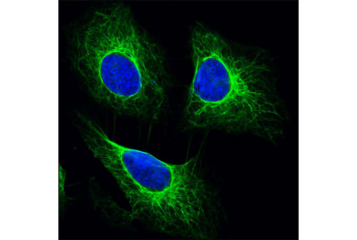
| 免疫组织化学(石蜡) | 1:500 - 1:2000 |
| 免疫荧光法(免疫细胞化学) | 1:100 - 1:400 |
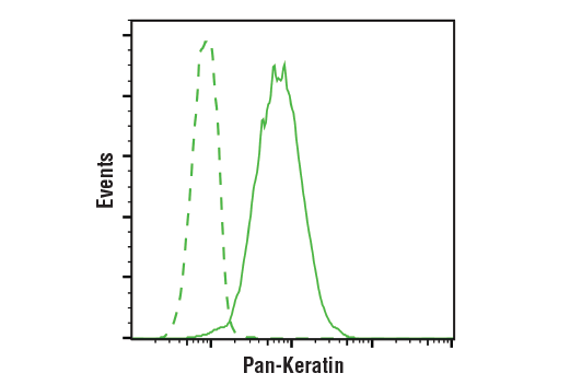
| 流式细胞术(固定/通透) | 1:200 - 1:800 |
蛋白质印迹法就是将膜与 5% w/v 脱脂奶粉、1X TBS 以及 0.1% Tween® 20 中的稀释的一抗在 4°C 下进行夜间孵育,同时轻轻晃动。
注意:请参阅一抗产品网页了解推荐的抗体稀释度。
从样品制备至检测,进行蛋白质印迹所需要的试剂现归于一个便利的试剂盒:#12957 Western Blotting Application Solutions Kit
注:使用反渗透去离子水 (RODI) 或同等级别的水配制溶液。
上样 20 µl 到 SDS-PAGE 凝胶 (10 cm x 10 cm) 上。
注意:建议将预染蛋白分子量标准品(#59329,10 µl/泳道)上样,以验证转膜的效率,生物素化蛋白质标准品(#7727,10 µl/泳道)可以直接在膜上显出条带以确定分子量。
注意:体积适用于 10 cm x 10 cm (100 cm2) 的膜;对于不同尺寸的膜,可相应调整体积。
* 避免反复接触皮肤。
发布时间 2005 年 6 月
修订时间 2020 年 6 月
实验步骤编号:19
注:使用反渗透去离子水 (RODI) 或同等级别的水配制溶液。
注意:在制作过程中务必随时保持切片处于湿润状态。
对于柠檬酸盐:将切片浸入 1 X 柠檬酸盐修复液,再放入微波炉中加热直至沸腾;继续保持亚沸腾温度 10 分钟 (95°-98°C)。在实验台上冷却切片 30 分钟。
|
推荐的 检测试剂 |
SignalStain® Boost IHC Detection Reagent (HRP, Mouse) #8125 | SignalStain® Boost IHC Detection Reagent (AP, Mouse) #31926 |
|---|---|---|
|
兼容的 色原 |
SignalStain® DAB Substrate Kit #8059 | SignalStain® Vibrant Red Alkaline Phosphatase Substrate Kit #76713 |
| SignalStain® Vivid Purple Peroxidase Substrate Kit #96632 | SignalStain® Ultra Blue Alkaline Phosphatase Substrate Kit #12824 | |
| SignalStain® Deep Black Peroxidase Substrate Kit #72986 | ||
| SignalStain® Radiant Yellow Peroxidase Substrate Kit #69644 |
注意:使用本实验步骤中未指定的检测试剂可能需要进一步优化一抗,考虑到检测试剂的不同灵敏度。
发布时间 2010 年 2 月
修订时间 2020 年 6 月
实验步骤编号:280
注意:用反渗透去离子 (RODI) 水或等效净化水制备溶液。
建议使用荧光物质标记的 Anti-Mouse 二抗:
注意:细胞应当在多孔板、腔室玻片或盖玻片上直接培养、处理、固定和染色。
注意:所有随后的孵育都应当在室温下完成,除非另有注明需要在避光湿盒或带盖的培养皿/板中孵育,以防止干燥和荧光物质淬灭。
发布时间 2010 年 12 月
实验步骤编号:145
本实验步骤中所需的所有试剂可与我们的 Intracellular Flow Cytometry Kit (Methanol) #13593 一起高效购买,或使用下文所列的货号单独购买。
注:使用反渗透去离子水 (RODI) 或同等级别的水配制溶液。
注:在您的实验中加入荧光细胞染料(包括活力指示染料、DNA 染料等)时,请参考染料产品网页,了解建议的实验步骤。访问 www.cellsignal.com,了解经验证用于流式细胞术的细胞染料完整列表。
注:固定前,贴壁细胞或组织应予以解离并处于单细胞悬液中。
注:最佳离心条件会根据细胞类型和试剂容量变动。一般,1-5 分钟 150-300g 将足以使细胞沉淀下来。
注:如果使用全血,则在固定前裂解红细胞并通过离心来洗涤。
注:如果表位被甲醛和/或甲醇破坏,可以在固定前添加靶向 CD 标记物或其他胞外蛋白质的抗体。在固定和透化过程期间,这类抗体将保持与目的靶标结合。但注意,某些荧光团(包括 PE 和 APC)会被甲醇损坏,因此不应在透化前添加。如果您不确定,请进行小规模实验。
注:使用血细胞计数器或备选方法计数细胞。
发布时间 2005 年 6 月
修订时间 2020 年 6 月
实验步骤编号:406
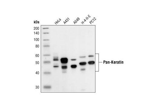
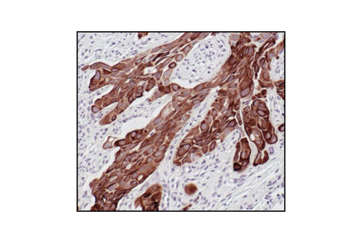
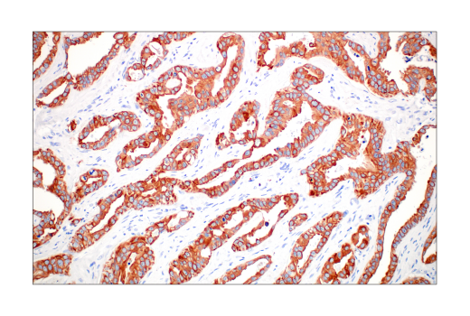
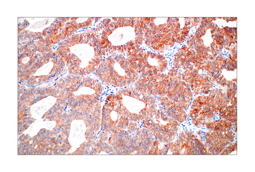
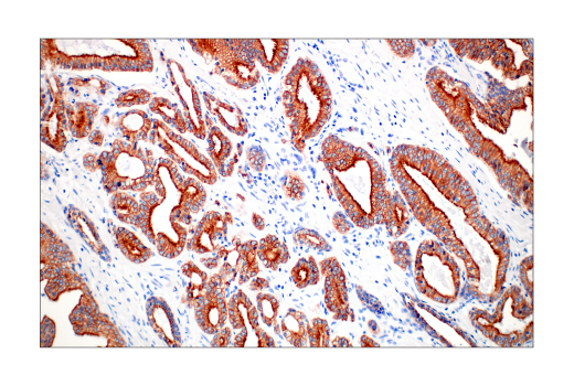
人, 小鼠, 大鼠, 猴
使用 A431 细胞的细胞骨架制剂对 BALB/c 小鼠进行免疫接种来产生单克隆抗体(同种型:IgG1)。
角蛋白(细胞角蛋白)是主要在上皮细胞中表达的中间丝蛋白。Keratin heterodimers composed of an acidic keratin (or type I keratin, keratins K9-K28) and a basic keratin (or type II keratin, keratins K1-K8 and K71-K80) assemble to form filaments. Keratin isoforms demonstrate tissue- and differentiation-specific profiles that make them useful as research and clinical biomarkers (1,2).
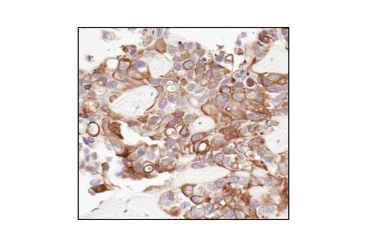
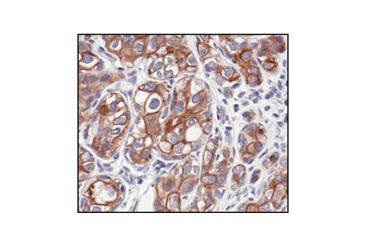
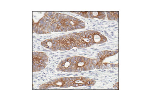
Dysregulation/mutations in keratin genes can lead to a variety of disorders affecting the skin, hair, nails, and other epithelial tissues (3). While expression of keratins can be variable, immunohistochemical staining of keratins is widely used to help in the identification and classification of epithelial tumors, and may also provide prognostic information.
Keratins 8 and 18 (K8/K18) are expressed in simple epithelia of normal tissue, as well as in adenocarcinomas of the breast, lung, ovary, and gastrointestinal tract. Keratin 17 is expressed in basal keratinocytes of stratified epithelia, hair follicles, and sebaceous glands. Onset of keratin 17 expression coincides with the definition of major epithelial lineages during skin development (4). Keratin 14 (K14) is expressed in basal cells of stratified epithelia, and in basal-like subtypes of breast cancer and squamous cell carcinomas. Keratin 19 (K19) is expressed in glandular epithelia, including the liver, gallbladder, and pancreas, as well as in adenocarcinomas of the breast, thyroid, and bile duct. Keratin 20 (K20) is expressed in gastrointestinal epithelium, urothelium, and Merkel cells in the skin, as well as in colorectal carcinomas and some urothelial carcinomas. Keratin 5/6 (K5/6) is expressed in basal cells of stratified epithelia, including the skin, prostate, and breast, as well as in basal-like breast cancers, squamous cell carcinomas, and some lung carcinomas. Keratin 7 (K7) is expressed in glandular epithelia, such as those in the lung, breast, and female reproductive tract, as well as in adenocarcinomas of the lung, breast, and ovary (5,6).
Keratins, particularly K8, K18, and K19, serve as biomarkers for identification of circulating tumor cells (CTCs) (5).
Post-translational modifications, including phosphorylation, acetylation, ubiquitylation, sumoylation, glycosylation, and transamidation, have been shown to affect the functions of keratins in normal and disease states (6). Understanding the molecular mechanisms underlying these PTMs may provide insights into cancer pathogenesis.
除非如以 CST 合法授权代表签署的书面形式另行明确同意,否则以下条款适用于 CST、其附属公司或其分销商提供的产品。除非 CST 合法授权代表以书面形式单独接受,否则任何附加于或异于此处所载条款和条件的客户条款和条件均被拒绝且无效。
产品用“仅供研究使用”或类似标示声明标示,并且尚未经 FDA 或其他国外或国内监管实体出于任何目的批准、准许或许可。客户不得出于任何诊断或治疗目的或以任何与产品标示声明相冲突的方式使用任何产品。CST 销售或许可的产品提供给作为最终用户的客户,且仅用于研究和开发用途。出于诊断、预防或治疗目的任何产品使用或出于转售(单独或作为成分)或其他商业目的的任何产品购买都要求来自 CST 的单独许可。客户 (a) 不得向任何第三方出售、许可、出借、捐赠或另行转让或提供任何本公司产品,无论单独或联合其他材料方式,或使用本公司产品制造任何商业产品,(b) 不得复制、修改、逆向工程、反编译、反汇编或另行尝试发现本公司产品的底层结构或技术,或出于开发与 CST 产品或服务竞争的任何产品或服务的目的使用本公司产品,(c) 不得从本公司产品改变或移除任何商标、商品名称、徽标、专利或版权声明或标记,(d) 仅应根据 CST 产品销售条款和任何适用文档使用本公司产品,以及 (e) 应就客户联系本公司产品所用的任何第三方产品或服务而言遵守任何许可、服务条款或类似协议。